Description
One attraction of the clinical discipline of hematology has been the ability to diagnose blood cell disorders quickly and relatively simply using tests that can be performed in a hematologist’s office: notably, blood cell counts, blood films, the reticulocyte count, and film of the marrow aspirate. Now these procedures are often performed in clinical laboratories, most with automated techniques. Nevertheless a physician, experienced and competent in the interpretation of blood counts and blood film, can determine the diagnosis or can reach a very limited differential diagnosis, quickly, in many cases.
Despite the increased specificity of hematological diagnosis provided by important ancillary analytical techniques, in many cases the blood counts and the blood and marrow films still permit the physician, expert in hematology, to deduce much of the information required to reach a diagnosis and to plan therapy. This initial diagnostic consideration can be supplemented by the important, indeed sometimes vital, additional testing available. Nevertheless, the presence of polychromatophilic macrocytes (reticulocytes) and characteristic red cell shape changes on the blood film may point to the specific type of hemolytic anemia, the white cell count and the differential count may be diagnostic of one of several types of leukemia, the platelet prevalence and morphology may be indicative of any of several thrombocytic disorders, and innumerable other diseases may be detected with the blood or marrow film, e.g., myeloma, lymphoma, Gaucher disease, Chediak-Higashi disease, and many others. A number of infectious diseases may be evident by the organisms parasitizing red or white cells. Numerous such examples are represented in this Atlas
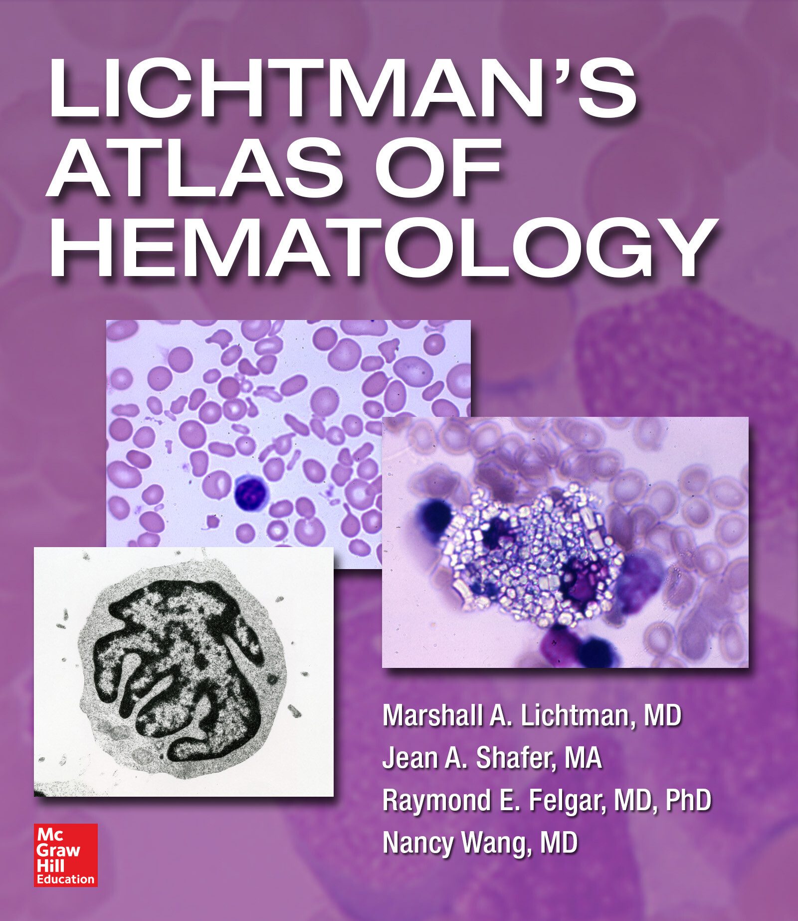
The intent of this work is to provide on-line access to images that are characteristic of a wide array of hematological disorders and other disorders that affect the blood, enhancing written descriptions found in Williams Hematology and other relevant medical books published on-line by McGraw-Hill. This Atlas permits an extensive presentation of blood, marrow, lymph node, spleen, and other organ pathology that would be too costly to be included in the print version of the text.
PART I: Red Cells I.A: Size, Chromicity, and Shape: Normal and Abnormal I.B: Pathological Red Cell Inclusions I.C: Red Cell Alterations in Non-Clonal Hematological Disorders PART II: White Cells II.A: Neutrophils, Normal II.B: Neutrophilia II.C: Neutrophils, Non-Clonal Abnormalities II.D: Eosinophils II.E: Basophil II.F: Monocytes and Macrophages II.G: Lymphocytes and Plasma Cells II.H: White Cell Concentrate (Buffy Coat) II.I: Carcinoma Cells in the Blood (Carcinocythemia) II.J: Leukoerythroblastic Reaction II.K: Other Non-Hematopoietic Cells in Blood (new section) PART III: Microorganisms III.A: Blood, Marrow, and Pleural Fluid PART IV: Platelets IV.A: Normal and Abnormal PART V: Marrow V.A: Cellularity V.B: Iron Stores V.C: Erythroid Precursors V.D: Myeloid Precursors V.E: Megakaryocytes V.F: Endothelial, Epithelial, Mast, Osteoblast and Osteoclast Cells V.G: Abnormal Erythroid Precursor Cells V.H: Normal & Abnormal Monocytes & Macrophages V.I: Lymphocytes and Plasma Cells V.J: Metastatic Tumor Cells V.K: Other Marrow Disorders V.L: Mitotic Figures PART VI: Clonal Myeloid Diseases VI.A: Acute and Chronic Leukemias VI.B: Clonal Mast Cell Diseases VI.C: Histiocytic and Myeloid Dendritic Leukemias PART VII: Lymphomas, Myeloma, and Lymphoid Leukemias VII.A: Normal and Reactive Lymph Node, Thymus, and Spleen VII.B: Precursor Lymphoblastic Lymphoma VII.C: Mature B-Cell Lymphomas and Myelomas VII.D: Mature T-Cell and NK-Cell Lymphomas VII.E: Hodgkin Lymphoma VII.F: Post-Transplant Lymphoproliferative Disorder VII.G: Lymphocytic Leukemias PART VIII: Cytogenetic Patterns VIII.A: Normal Karyotype VIII.B: Acute Myelogenous Leukemia VIII.C: Chronic Myelogenous Leukemia VIII.D: Acute Lymphocytic Leukemia VIII.E: Chronic Lymphocytic Leukemia VIII.F: Lymphoma VIII.G: Myeloma VIII.H: Special Techniques of Chromosome Identification PART IX: Flow Cytometric Patterns IX.A: Normal Blood, Marrow, and Lymph Node Cells IX.B: Acute Myelogenous Leukemia IX.C: Lymphocytic Leukemia and Lymphoma IX.D: Plasma Cell Neoplasms IX.E: Paroxysmal Nocturnal Hemoglobinuria (PNH) PART X: Cytochemistry and Immunostains X.A: Peroxidase Stain X.B: Esterase Stains X.C: Immunostains X.D: In Situ Hybridization Methods X.E: BCL-2 and ALK-1 Immunostains X.F: Special Stains for Mast Cells and Langerhans Cells X.G: Congo Red Stain for Amyloid Protein X.H: Toluidine Blue Stain: Mast Cells and Basophils X.I: Glycoprotein IIIa Stain: Platelets and Megakaryocytes X.J: von Willebrand Factor Immunofluorescence X.K: Leukocyte Alkaline Phosphatase X.L: Acid Phosphatase X.M: Sudan Black B Stain PART XI: External Manifestations XI.A: External Manifestations PART XII: Marrow Ultrastructure and Cell Egress XII.A: Marrow Circulation XII.B: Marrow Hematopoietic Cord and Sinus Structure XII.C: Erythrocyte Egress XII.D: Granulocyte Egress XII.E: Mononuclear Leukocyte Egress XII.F: Proplatelet Egress XII.G: Neural


