Description
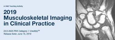
The CME Activity provides a review of clinical applications concerning the diagnosis, treatment and management of the musculoskeletal disorders. Modern imaging strategies, surgical correlation and the need for intra-disciplinary teamwork when deciding the most effective patient management will be addressed. Faculty share techniques and tips in image interpretation of musculoskeletal injuries and pathology. Information gained during this activity is geared to improve diagnostic capabilities and expand clinical applications. The faculty has been selected for their teaching ability, as well as for their expertise.
Target Audience
This CME activity is designed to educate diagnostic imaging physicians who supervise and interpret musculoskeletal MRI. In addition, referring physicians who order musculoskeletal MRI should gain an appreciation of the strengths and limitations of these types of procedures.
Educational Objectives
At the completion of this CME teaching activity, you should be able to:
- Incorporate state of the art imaging protocols to accurately assess musculoskeletal pathology and injury.
- Assess patients with joint pathology in a non-invasive manner utilizing MRI.
- Recognize the strengths and limitations of MRI for the management of sports related injuries.
- Describe the MR appearance of muscle and tendon injury.
- Differentiate musculoskeletal masses and tumors.
- Modify image sequencing to accurately evaluate joint replacement and minimize artifact.
- Discuss the utility of imaging when used to guide therapeutic musculoskeletal injections.
Topics And Speakers:
Session 1
MRI of the Knee Menisci: Tricks and Tips
William B. Morrison, M.D.
MRI of the Knee: Ligaments and Miscellaneous Disorders
Mark H. Awh, M.D.
MRI of the Knee: Case Based
John F. Feller, M.D.
Session 2
Posterolateral Corner of the Knee Made Simple
William B. Morrison, M.D.
MRI of the Knee: Arthroscopic Validation of MRI Findings
Matthew Diltz, M.D.
MRI of the Foot and Ankle
Mark H. Awh, M.D.
Session 3
Femoroacetabular Impingement Update
Geoffrey M. Riley, M.D.
Current Concepts in Imaging and Arthroscopy of the Hip
Matthew Diltz, M.D.
Session 4
MRI and Bone Marrow; Practical Solutions to Common Problems
Geoffrey M. Riley, M.D.
Degenerative Disc Disease, Pathophysiology and Nomenclature
Jeffrey S. Ross, M.D.
Session 5
The Craniocervical Junction
Jeffrey S. Ross, M.D.
What Level Is This: Making Sense of Transitional Spinal Anatomy
Geoffrey M. Riley, M.D.
MRI of Spine Infection and Differential Diagnosis
William B. Morrison, M.D.
Session 6
MR of Spine Trauma
Jeffrey S. Ross, M.D.
Pearls and Pitfalls in Shoulder MRI
Lynne S. Steinbach, M.D.
Postoperative Spine Imaging
Jeffrey S. Ross, M.D.
Session 7
The Throwing Shoulder
Lynne S. Steinbach, M.D.
Recent Articles Important to Your Practice
Geoffrey M. Riley, M.D.
MR of Sports Injuries in the Professional Athlete
Mark H. Awh, M.D.
Session 8
Can We Determine Return to Play?
William B Morrison, M.D.
MRI Following Joint Replacement
John F. Feller, M.D.
MRI of Musculoskeletal Masses and Tumor-like Conditions
Mark H. Awh, M.D.
Session 9
Biceps Muscle and Tendon in the Upper Extremity
Lynne S. Steinbach, M.D.
Bone, Tendon and Muscle Injuries
Matthew Diltz, M.D.
Image Guided Diagnostic and Therapeutic MSK Injections
John F. Feller, M.D.
Session 10
Postoperative Shoulder
Lynne S. Steinbach, M.D.
Orthopedic Injuries: From Diagnosis to Treatment
Matthew Diltz, M.D.
Session 11
New & Proven Techniques for Rapid Musculoskeletal MRI
Jan Fritz, M.D., P.D., D.A.B.R.
Automated MR Imaging of the Musculoskeletal System
John F. Feller, M.D.
Metal Artifacts in MRI: Practical Problem Solving
Jan Fritz, M.D., P.D., D.A.B.R.
Session 12
MRI of the Shoulder
John F. Feller, M.D.
Elbow MRI
Jan Fritz, M.D., P.D., D.A.B.R.
MRI of the Hip
John F. Feller, M.D.
Multi-Detector CTA of Extremity Trauma
Jan Fritz, M.D., P.D., D.A.B.R.


Avis
Il n’y a pas encore d’avis.