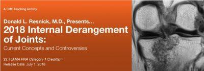
Internal Derangement of Joints: Current Concepts and Controversies 2018 (Videos) Regular price
Format: 31 Video Files (.mp4 format).
$26.99
About This CME Teaching Activity
This CME activity provides an update of current information on MR Imaging in the assessment of musculoskeletal disorders. Essential anatomy, physiology and pathology are emphasized that explain imaging findings in disorders of the shoulder, elbow, wrist, hand, hip, knee, ankle and foot. MR imaging findings in the assessment of common problems in peripheral joints are compared to those derived from other imaging methods.
Educational Objectives
At the completion of this CME teaching activity, you should be able to:
Assess MR images in patients with internal derangements of peripheral joints.
Articulate the anatomic features fundamental to accurate interpretation of the MR imaging findings in these disorders.
Formulate a reasonable differential diagnostic list and be able to identify the most likely diagnosis.
Comprehend the pathogenesis and imaging findings associated with common and important disorders that affect all joints.
Topics And Speakers:
Session 1
Common and Uncommon Disorders of Synovium-Lined Joints
Donald L. Resnick, M.D.
Intra-Articular Tumors and Tumor-Like Processes
Edward Smitaman, M.D.
Session 2
Osteochondral, Subchondral, Cortical: Acute Injuries
Donald L. Resnick, M.D.
Osteochondral, Subchondral, Cortical: Cartilage Imaging
Christine B. Chung, M.D.
Osteochondral, Subchondral, Cortical: Stress Injuries
Mini N. Pathria, M.D.
Session 3
Pectoralis Major/Minor and Teres Major/Latissimus Dorsi Tendons
Eric Y. Chang, M.D.
Rotator Cuff: Basic and Footprint Anatomy and Patterns of Failure with Emphasis on Terminology, Tears and Pseudotears, Impingement, and Calcification
Donald L. Resnick, M.D.
Biceps Tendon and Rotator Interval
Eric Y. Chang, M.D.
Session 4
Entrapment Neuropathies of the Upper Extremity
Evelyne A. Fliszar, M.D.
The Brachial Plexus
Brady Huang, M.D.
Shoulder Superior Labrum: Anatomy, Normal Variants, SLAP Lesions, and Microinstability
Donald L. Resnick, M.D.
Session 5
Macroinstability of the Glenohumeral Joint Including Various Patterns of Failure of the Labrum and Glenohumeral Ligaments
Donald L. Resnick, M.D.
Throwing Shoulder
Donald L. Resnick, M.D.
The Acromioclavicular Joint
Mini N. Pathria, M.D.
Session 6
Muscle Disorders
Mini N. Pathria, M.D.
Elbow: Anatomy, Ligaments, Instability, and the "Throwing" Elbow
Donald L. Resnick, M.D.
Session 7
Elbow Tendon Anatomy and Pathology with Emphasis on the Biceps, Brachialis, Triceps, and Flexor and Extensor Tendons
Donald L. Resnick, M.D.
Wrist Triangular Fibrocartilage Complex (TFCC): Anatomy and Patterns of Failure
Donald L. Resnick, M.D.
Wrist Intrinsic and Extrinsic Ligaments/Instability
Donald L. Resnick, M.D.
Session 8
Interesting Cases: Upper Extremity
Karen C. Chen, M.D.
Entrapment Neuropathies of the Lower Extremity
Tudor Hughes, M.D., FRCR
Hip Joint Anatomy and External and Internal Femoroacetabular Impingement
Donald L. Resnick, M.D.
Session 9
Knee Anatomy Biomechanics and Overview of Injury Patterns
Donald L. Resnick, M.D.
Meniscus: Anatomy and Patterns of Failure
Donald L. Resnick, M.D.
Session 10
Muscles and Tendons about the Hip: Anatomy, Strains, and Tears
Mini N. Pathria, M.D.
Knee: Extensor Mechanism
Mini N. Pathria, M.D.
Knee: Postoperative Ligaments
Brady Huang, M.D.
Session 11
Knee: Cruciate Ligaments
Brady Huang, M.D.
Knee: Medial and Lateral Supporting Structures
Brady Huang, M.D.
Session 12
Ankle Tendons: Normal Anatomy, Tendinosis, Tenosynovitis, and Tendon Tears
Donald L. Resnick, M.D.
Ankle Ligaments: Normal Anatomy and Patterns of Injury
Donald L. Resnick, M.D.
Showing 1–12 of 1220 results
-
Sale!

Orthopaedic Pathology Third Edition
$19.99 Add to cart -
Sale!
Textbook of Orthopaedics 3rd Edition
$12.99 Add to cart -

Periprosthetic Joint Infection: Updated Practical Management Guide 2nd Edition
$9.99 Add to cart -
Sale!
Cervical Myelopathy 1st Edition
$11.99 Add to cart -
Postgraduate Orthopedics: An Exam Preparatory Manual 3rd Edition
$8.99 Add to cart -
Sale!

Turek’s Orthopaedics Principles & Their Applications 8th Edition 2 Volume Set
$27.99 Add to cart -
Sale!

Orthopaedic Knowledge Update®: Shoulder and Elbow 6th Edition
$27.99 Add to cart -
Sale!

Orthopaedic Knowledge Update®: Musculoskeletal Tumors 5: OKU Fifth Edition
$27.99 Add to cart -
Sale!

Workbook for Diagnostic Medical Sonography: Obstetrics and Gynecology 5th edition
$8.99 Add to cart -

Biochemistry for Sport and Exercise Metabolism Second Edition
$11.99 Add to cart -
Energetic Kinesiology: Principles and Practice 1st Edition
$9.99 Add to cart -

At the Still Point of the Turning World The Art and Philosophy of Osteopathy 1st Edition
$11.99 Add to cart
